Wednesday, November 15, 2023

Aspirin, Overview, Indications, Contraindications & Side effects

Aspirin, Overview, Indications, Contraindications & Side effects  

Overview:

Aspirin is an NSAID (non-steroidal anti-inflammatory drug). It has anti-infalammatory, analgesic, antipyretic and antithrombotic activity. It interferes with prostaglandin synthesis by irreversibly inhibiting cyclooxygenase. It was synthesized in 1853, but the drug was not in use until 1899, when it was found to be effective in the arthritis and well tolerated. The name it was coined from the German word for the compound, acetylspirsaure. Because of its greater efficacy and lower cost, it rapidly replaced the natural products in use at that time, and has remained one the most widely employed remedies for over 90 years.

Indications:

Aspirin is primarily indicated in conditions like Fever, Following coronary bypass surgery, Headache, Inflammation, Ischaemic event (unstable angina, myocardial infarction), Mild to moderate pain, Myocardial infarction, Myocardial infarction prophylaxis, Pain and inflammation (in rheumatic disease, in other musculoskeletal disorders), Prophylaxis of cerebrovascular disease, Pyrexia, Stable angina, Stable angina pectoris, Stroke prophylaxis, and can also be given in adjunctive therapy as an alternative drug of choice in Arterial thromboembolism prophylaxis, Colorectal carcinoma prophylaxis, Juvenile rheumatoid arthritis, Neurogenic bladder, Osteoarthritis.

Contraindications:

Aspirin is contraindicated in conditions like Gl ulceration, Hypoprothrombinemia, Hypersensitivity.

Side Effects:

The severe or irreversible adverse effects of Aspirin, which give rise to further complications include Urticaria, Gi bleeding, Rhinitis, Angioneurotic edema, Hepatitis, Hepatomegaly.Aspirin produces potentially life-threatening effects which include Cerebral hemorrhage, Airway obstruction, Gastric ulceration, Gastric erosion. which are responsible for the discontinuation of Aspirin therapy.The signs and symptoms that are produced after the acute overdosage of Aspirin include Coma, Acidosis, Hyperthermia, Volume depletion, Hyperglycemia, Renal failure, Hyperventilation, Cardiovascular collapse, Vertigo, Tinnitus, Hyperkalemia.The symptomatic adverse reactions produced by Aspirin are more or less tolerable and if they become severe, they can be treated symptomatically, these include Dizziness, Vertigo, Nausea, Vomiting, Tinnitus, Epigastric discomfort, Dyspepsia, Asthma, Respiratory alkalosis, Bleeding.

Warnings:

Aspirin should be used with caution in patients with chronic renal insufficiency, gastric ulcer, severe anemia, and patients intolerant to salicylate. Avoid aspirin in patient with history of blood coagulation defects and if patient taking anticoagulants. Liver function should be monitored in patients receiving large doses of aspirin or in patients with pre-existing hepatic disease in order to prevent reversible, dose dependent hepatotoxicity.

High Risk Groups:

Drug should not be given to Paediatrics, Pregnant Mothers, patients suffering from Kidney dysfunction, patients suffering from Liver Malfunction, and Neonates.If prescribing authority justifies the benefits of the drug against the possible damages he/she should reevaluate them and consult the reference material and previous studies.

Pregnancy:

Avoid chronic or intermittent high doses during pregnancy; may affect maternal and newborn hemostasis mechanisms, leading to an increased risk of hemorrhage.

Premature closure of the ductus arteriosus may occur if used near term with use of full-dose aspirin.


Acetaminophen, Paracetamol, Overview, Indications, Contraindications & Side effects

 Acetaminophen, Paracetamol, Overview, Indications, Contraindications & Side effects 

Overview:

Paracetamol is analgesic and antipyretic agent. Paracetamol is the active metabolite of phenacetin, responsible for its analgesic effect. Paracetamol is a weak prostaglandin inhibitor in peripheral tissues and possesses no significant antinflammatory effects. Paracetamol is one of the most important drug used for the treatment of mild to moderate pain when an antinfalmmatory effect is not necessary. Paracetamol is preferred over aspirin as an analgesic/antipyretic for patients in whom aspirin is contraindicated, such as those who have a history of gastric ulcer or a coagulation disorder.

Indications:

Paracetamol is primarily indicated in conditions like Ear pain, Fever, Headache, Malaise, Migraine, Mild to moderate pain, Pain, Post-vaccine reaction, Short-bowel syndrome, Tobacco amblyopia and leber's optic atrophy, Toothache.

Contraindication:

Paracetamol is contraindicated in conditions like Hypersensitivity.

Side Effects:

The severe or irreversible adverse effects of Paracetamol, which give rise to further complications include Bronchospasm.Paracetamol produces potentially life-threatening effects which include Blood dyscrasias, Centribular Necrosis, Liver damage. which are responsible for the discontinuation of Paracetamol therapy. The signs and symptoms that are produced after the acute overdosage of Paracetamol include hypoglycemic coma, Hepatic necrosis, Liver failure, renal tubular necrosis.The symptomatic adverse reactions produced by Paracetamol are more or less tolerable and if they become severe, they can be treated symptomatically, these include Skin rashes, Gl adverse effects.

Warnings:

If sensitivity reaction occurs, discontinue use of paracetamol. If pain persist more than 10 days and arthritic and rheumatic condition affecting children, immediately consult physician. If patient have been diagnosed with liver or kidney impairment, seek medical advice before taking medication. If symptoms persists consult doctor.

Pregnancy:

Drug crosses placenta and can be detected in cord blood, newborn serum, and urine immediately after delivery.
Increased risk of teratogenic effects not reported.
Use of normal doses during pregnancy not associated with increased risk of miscarriage or still birth; however, increase in fetal death or spontaneous abortion may be seen with maternal overdose if treatment delayed.


Sunday, November 12, 2023

Oral Infection

 Oral Infection:

Overview:

Infection involving the oral cavity can be associated with significant morbidity. In addition, recent studies have suggested that some types of oral infection may potentially confound a number of systemic problems including cardiac disease, pregnancy, kidney disease, and diabetes. 

Relevant anatomy:

The oral cavity (see the image below) is oval shaped and is separated into the oral vestibule and the oral cavity proper. It is bound by the lips anteriorly, the cheeks laterally, the floor of the mouth inferiorly, the oropharynx posteriorly, and the palate superiorly. The oropharynx begins superiorly at the junction between the hard palate and the soft palate, and inferiorly behind the circumvallate papillae of the tongue. The bony base of the oral cavity is represented by the maxillary and mandibular bones.

The tongue is basically a mass of muscle that is almost completely covered by a mucous membrane. It occupies most of the oral cavity and oropharynx. It is known for its role in taste, but it also assists with mastication (chewing), deglutition (swallowing), articulation (speech), and oral cleaning. Five cranial nerves contribute to the complex innervation of this multifunctional organ.

Microbiology of Oral Infection:

Oral bacteria are diverse, but in the immunocompetent individual the interaction between the various complex competing microflora provides relative stability. The different structures and anatomy within the mouth afford various microenvironments that support different types of microbiologic organisms. In the first few years of life, the bacterial microbiota within the mouth is predominantly aerobic, but, as the teeth develop, favorable sites supporting pathogenic anaerobic bacteria emerge.

Biofilm, on teeth termed plaque, can build up in the mouth and reach substantial numbers. It has been estimated to reach up to 1011 microorganisms/mL in plaque that is not removed over several days. Apical periodontal infection has been associated with 200 bacterial species, and 500 bacterial species have been reported with marginal periodontitis. Evidence also exists that significant interaction of bacterial types within biofilm may either enhance or suppress metabolic activity that leads to dental infection.

Factors that regulate the number of oral bacteria include general exposure, saliva, diet, bacterial retention, bacterial interaction, the complexity of the flora, native resistance, and hygiene. Change in the condition of any one of the above factors or combination thereof can result in oral hard or soft tissue infection.

For example, a diet rich in dietary carbohydrate such as refined sugar favors bacteria such as Streptococcus mutans, the organism that causes dental caries. The consistency of the diet is also important in that courser foods eliminate particles that can sustain microorganisms. Specificity in terms of bacteria tissue adherence also appears to exist; for example, S mutans and Streptococcus sanguis typically adhere to hard surfaces, while Streptococcus salivarius is found primarily on the tongue.

A number of systemic diseases can reduce host defense mechanisms, leading to reductions in phagocytic activity, pulmonary clearance, and circulation, with these factors contributing to oral infection. They include malnutrition, alcoholism, diabetes, cystic fibrosis, renal failure, lymphoproliferative disorders, heart failure, and chronic lung disease.

Immunosuppressants and other medications used as cytotoxic chemotherapeutic agents, including steroids, can reduce host defense mechanisms leading to infection. Antibiotic medication can also alter the oral microbial environment leading to oral infection. For example, prolonged antibiotic therapy can reduce the normal bacterial flora, resulting in the selection of resistant flora and/or the emergence of competing fungal organisms. Other factors potentially confounding the possibility of oral infection include age, potential drug abuse, family and social environment, and the patient’s psychological status.

Oral infection can also be caused by viral and fungal organisms. Common viral infections include herpes simplex (primary and secondary recurrent herpes infection), varicella zoster (shingles), and coxsackievirus. The presence of the papilloma virus is considered an infection and has assumed importance in that it has been linked to the development of oral squamous cell carcinomas.

Viral and Fungal Oral Infection:

As mentioned previously, oral infection can also be caused by viral (eg, herpes simplex [primary and secondary recurrent herpes infection], varicella zoster [shingles], coxsackievirus) and fungal organisms. The presence of the papilloma virus (see image below) is considered an infection and has assumed importance in that it has been linked to the development of oral squamous cell carcinomas. 


Primary viral infections involving the oral mucosa are covered elsewhere. However, a condition not typically described in the literature is intraoral recurrent herpes (see image below). This infectious process is the equivalent of the secondary recurrent lesion of herpes but occurs on the attached gingival tissue adjacent to the teeth, including the palate, rather than the lip. A prodromal phase often exists in which the tissue feels itchy or tingles. This dysesthesia is then followed by the development of multiple small punctuate ulcers that coalesce and are painful. The infection may last 10-14 days and reoccur in response to stress, fever, trauma, and hormonal alterations. No good evidence exists that sunlight activates the latent virus, as occurs with the secondary lip lesion.


The most common oral mycotic infection is candidiasis, but other infections such as mucormycosis (phycomycosis), actinomycosis (see image below), aspergillosis, and pneumocystosis can occur under the right circumstances. Candida is an opportunistic organism and does not typically cause infection unless other host factors are compromised. Systemic conditions that can contribute to candidiasis include xerostomia, diabetes mellitus, and immune system suppression (eg, HIV infection or as a side effect of drying medications). Candidiasis can also result from poorly fitting prosthetic oral appliances and poor oral hygiene.

The most common oral mycotic infection is candidiasis, but other infections such as mucormycosis (phycomycosis), actinomycosis (see image below), aspergillosis, and pneumocystosis can occur under the right circumstances. Candida is an opportunistic organism and does not typically cause infection unless other host factors are compromised. Systemic conditions that can contribute to candidiasis include xerostomia, diabetes mellitus, and immune system suppression (eg, HIV infection or as a side effect of drying medications). Candidiasis can also result from poorly fitting prosthetic oral appliances and poor oral hygiene.

Candidiasis can occur in 3 forms: pseudomembranous, atrophic/erythematous, and chronic/hyperplastic. The first form, pseudomembranous, is characterized by multiple soft, white, slightly elevated plaques that can be wiped away, leaving an erythematous and bleeding surface (see image below). The second form is characterized by erythematous tissue that is sensitive to touch, and the third by confluent slightly raised white areas. Culturing for candida is useful in cases in which the diagnosis is unclear. 

Caries and Periapical Infection:

Caries are the most common odontogenic infection. Prevalence appears to vary depending on the region of the world surveyed. When dental infection is limited to the enamel and dentine of the tooth, it does not present a severe problem. A carious process that extends into the dental pulp chamber and the neural, vascular, and connective tissue contained within presents a more serious matter.

The bacteria that reach the pulp are mostly anaerobic and host resistance is limited by the constricted anatomy of the tooth pulpal chamber. Once the pulp has become involved, the inflammation and edema associated with the carious infection causes a vascular necrosis and the death of this tissue. This process then generally results in the infection spreading beyond the apex of the root (ie, the periapical region).

Osteomyelitis and Cellulitis:

Once the infectious process has extended beyond the tooth, it may expand into the surrounding medullary bone and cause an osteomyelitis, or it may extend focally through the bone as a fistular tract draining into the oral cavity. The latter condition drains the infection, reduces general swelling, and results in a small soft tissue swelling and focal erythema at the site of the mucosal involvement.

However, a periapical infection may also evolve into a localized soft tissue abscess or a more diffuse and troublesome soft tissue cellulitis. The spread of infection into fascial spaces can be problematic for the patient and treating clinician. A cellulitis can involve incorporation of adjacent spaces and inferior (eg, neck and mediastinal region) and superior (eg, base of the skull, foramen ovale, and brain — cavernous sinus thrombosis) extension.

The relative risk of fascial space infection is related to the location of the space in relation to the anatomy of the head and neck. Low-risk anatomical areas include the region of the facial vestibule of the mandible, the body of the mandible, the buccal vestibule of the mandible, and the palate. Moderate risk areas include the mentalis space, submental space, sublingual space, submandibular space, buccal space, submasseteric space, superficial and deep temporal space, and the maxillary sinus. The highest-risk regions include the infratemporal and parotid spaces; the pterygomandibular, parapharyngeal, and peritonsillar spaces; and the cervical, infraorbital, periorbital region, and the base of the lower lip. Infection that has spread to these latter areas must be treated aggressively.

Signs and symptoms for the various space infections vary. For example, infection of the sublingual space itself is not evident by external examination. However, the patient feels like the tongue has been pushed up, and difficulty swallowing can exist. In contrast, Ludwig angina, which represents a more extensive involvement, not only of the sublingual space but space below the mandible, severely displaces the tongue upwards and backwards. Extensive swelling of the anterior neck extending down to the clavicles may become evident. With extension of the infection, the patient is febrile and breathing is often labored. Obstruction of the throat due to edema of the glottis causes further deterioration.

Other commonly observed general signs of cellulitis include jaw-opening limitation (eg, trismus), and distension of the involved region (eg, the lip, cheek), and bulging of muscle (eg, the temporalis). In cases of parotid space infection, suppuration from the parotid duct may be present. Typically, pain is described as throbbing and severe tenderness to palpation exists. Pain worsens often quickly over time and is not controlled by nonprescription medication.

Osteomyelitis can follow chronic odontogenic infection not involving cellulites. The incidence of the disease has decreased with the advent of antibiotics, caries prevention, and restorative dentistry. However, chronic diseases, immune deficiency, and vascular abnormality may predispose an individual to the infection.

The course of osteomyelitis can include suppuration with abscess or fistula formation and jaw bone sequestration. Acute osteomyelitis is associated with the classical features of infection including fever, leukocytosis, lymphadenopathy, pain, and soft tissue swelling. If the condition involves the lower jaw there may be lip dysesthesia or paresthesia. Chronic osteomyelitis may also involve purulent discharge, tooth loss, or pathologic fracture. The condition typically involves symptom remission and recrudescence. Note that osteomyelitis can result from local or metastatic neoplasm as well as jaw bone fracture.

Diffuse sclerosing osteomyelitis is a variation of osteomyelitis, but it involves fairly focal areas of chronic infection that can cause periosteal and cortical bulging in adults (typically) without frank sequestration of the cortex. However, internal sequestration occurs. Low-grade persistent pain exists. Condensing osteitis is the name of the condition involving bone sclerosis associated with long-standing pulpitis or pulpal necrosis without demonstrative infection. Infection associated with removal of a tooth is termed alveolar osteitis or dry socket. Another newly defined condition involving infection of implants from adjacent dental infection is termed retrograde periimplantitis.

Periodontal Disease:

The most common oral mucosal infection is periodontal disease. This condition is considered a major public health problem in many countries.  Periodontitis has been associated with the proliferation of the bacteria Actinobacillus actinomycetemcomitans,Porphyromonas gingivalis, and Prevotella intermedia. Infection involves the supporting structures of the teeth and causes progressive loss of gingival attachment with absorption of supporting alveolar bone. This results in periodontal pocket formation, which can lead to further disease including tooth loss. When a deep periodontal pocket closes along the superior interface with a tooth trapping bacteria, this condition can lead to abscess.

Several forms of periodontal infection are described. They include aggressive periodontitis, which occurs in otherwise healthy individuals but causes rapid bone loss; chronic periodontitis characterized by gingival pocket formation and bone loss occurring over a prolonged period of time; periodontitis as a manifestation of systemic disease; and necrotizing periodontal disease, which involves severe necrosis of the gingival tissue. This condition is most often associated with the presence of systemic conditions such as HIV infection, malnutrition, and immunosuppression.

Known contributory factors to periodontal infection include age, plaque and calculus, smoking, diabetes mellitus, general health status, and social economic level. Low bone density of osteoporosis appears to be a shared risk factor for periodontitis rather than a causal factor. A study by Teeuw et al on 313 dental patients found that suspected new diabetes was found in 18.1% of patients diagnosed with severe periodontitis (78 patients) compared to 9.9% and 8.5% in patients with mild or moderate periodontitis and the control group.

Periodontitis is preceded by infection and inflammation of the attached gingival adjacent to teeth. This is termed gingivitis. Both gingivitis as well as periodontitis may be asymptomatic unless a frank abscess exists. Symptoms of gingivitis and periodontitis include foul breath and gingival tenderness with palpation; signs can include gingival bleeding, tooth mobility, and gingival erythema and swelling. 
Moderate chronic gingivitis. Note that the papillae are edematous and blunted. They may bleed with brushing. Note areas of edema overlying some of the root areas. Pallor is seen in these areas.

Severe periodontal disease. Loss of the gingival tissue is seen, making the teeth appear long. Even more effacement of the papillae is present. Heaped up ridges are observed in the areas overlying the roots.


Minor Salivary Gland Infection:


Infection involving the intraoral minor salivary glands is relatively rare. However, minor gland infection can follow sialolithiasis. This condition begins with the formation of a small calculi or stone that blocks the duct of the minor gland. A secondary infection may then ensue. Sialadenitis can also follow viral infection and can be associated with neoplastic activity. The condition typically causes erythema and swelling of the minor gland duct. Minor glands are located throughout the mouth but most frequently in the cheek, lip, and palatal region. Major salivary gland infection including acute and chronic bacterial parotitis, viral parotitis (ie, mumps), and other conditions causing salivary gland infection such as tuberculosis and parotid or submandibular actinomycosis is covered elsewhere.


Thursday, November 9, 2023

Alopecia Areata, Causes, Symptoms, Treatment

 Alopecia Areata:


Overview of Alopecia Areata:

Alopecia areata is a disease that happens when the immune system attacks hair follicles and causes hair loss. Hair follicles are the structures in skin that form hair. While hair can be lost from any part of the body, alopecia areata usually affects the head and face. Hair typically falls out in small, round patches about the size of a quarter, but in some cases, hair loss is more extensive. Most people with the disease are healthy and have no other symptoms.

The course of alopecia areata varies from person to person. Some have bouts of hair loss throughout their lives, while others only have one episode. Recovery is unpredictable too, with hair regrowing fully in some people but not others.

There is no cure for alopecia areata, but there are treatments that help hair grow back more quickly. There are also resources to help people cope with hair loss.

Types of Alopecia Areata:

There are three main types of alopecia areata:

Patchy alopecia areata: In this type, which is the most common, hair loss happens in one or more coin-sized patches on the scalp or other parts of the body.

Alopecia totalis: People with this type lose all or nearly all of the hair on their scalp.

Alopecia universalis: In this type, which is rare, there is a complete or nearly complete loss of hair on the scalp, face, and rest of the body.

Symptoms of Alopecia Areata:

Alopecia areata primarily affects hair, but in some cases, there are nail changes as well. People with the disease are usually healthy and have no other symptoms.

Hair Changes

Alopecia areata typically begins with sudden loss of round or oval patches of hair on the scalp, but any part of the body may be affected, such as the beard area in men, or the eyebrows or eyelashes. Around the edges of the patch, there are often short broken hairs or “exclamation point” hairs that are narrower at their base than their tip. There is usually no sign of a rash, redness, or scarring on the bare patches. Some people say they feel tingling, burning, or itching on patches of skin right before the hair falls out.

When a bare patch develops, it is hard to predict what will happen next. The possibilities include:

The hair regrows within a few months. It may look white or gray at first but may regain its natural color over time.
Additional bare patches develop. Sometimes hair regrows in the first patch while new bare patches are forming.
Small patches join to form larger ones. In rare cases, hair is eventually lost from the entire scalp, called alopecia totalis.
There is a progression to complete loss of body hair, a type of the disease called alopecia universalis. This is rare.
In most cases, the hair regrows, but there may be subsequent episodes of hair loss.

The hair tends to regrow on its own more fully in people with:

Less extensive hair loss.
Later age of onset.
No nail changes.
No family history of the disease.
Nail Changes

Nail changes such as ridges and pits occur in some people, especially those who have more extensive hair loss.

Causes of Alopecia Areata:

In alopecia areata, the immune system mistakenly attacks hair follicles, causing inflammation. Researchers do not fully understand what causes the immune attack on hair follicles, but they believe that both genetic and environmental (non-genetic) factors play a role.

Who Gets Alopecia Areata?

Anyone can have alopecia areata. Men and women get it equally, and it affects all racial and ethnic groups. The onset can be at any age, but most people get it in their teens, twenties, or thirties. When it occurs in children younger than age 10, it tends to be more extensive and progressive.
If you have a close family member with the disease, you may have a higher risk of getting it, but for many people, there is no family history. Scientists have linked a number of genes to the disease, which suggests that genetics play a role in alopecia areata. Many of the genes they have found are important for the functioning of the immune system.

People with certain autoimmune diseases, such as psoriasis, thyroid disease, or vitiligo, are more likely to get alopecia areata, as are those with allergic conditions such as hay fever.

It is possible that emotional stress or an illness can bring on alopecia areata in people who are at risk, but in most cases, there is no obvious trigger.

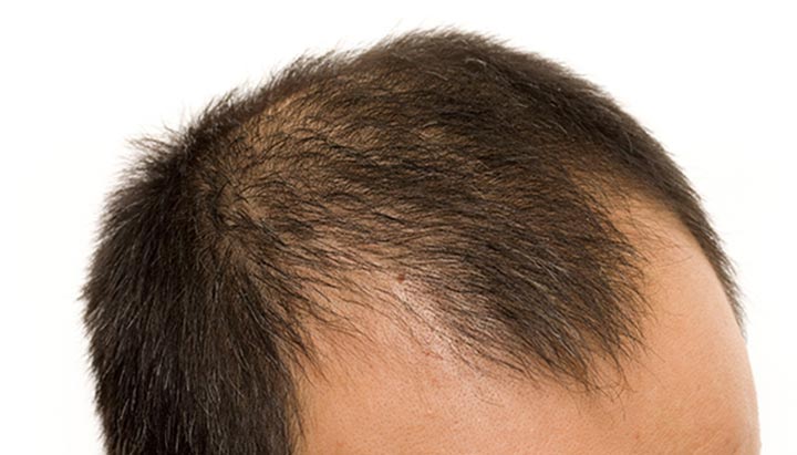
What is male pattern baldness?

Male pattern baldness (androgenic alopecia) is a type of hair loss that affects people assigned male at birth (AMAB). It causes you to lose hair on the skin covering your head (scalp), and your hair doesn’t grow back. Other signs of male-pattern baldness include thinning hair and a hairline that moves farther back on your head (receding hairline).

There are seven stages of male pattern baldness according to the Hamilton-Norwood scale:

Stage 1: There’s little or no hair loss or hairline recession.
Stage 2: There’s slight hair loss near the skin between your ears and forehead (temples).
Stage 3: You have deep hairline recession around your temples, and your hairline may have an “M” or “U” shape.
Stage 4: You have very deep hairline recession and a loss of hair at the top of your head (crown).
Stage 5: Your hairline recession connects to the bald spot on your crown.
Stage 6: The hair between your temples and crown is thinning or gone.
Stage 7: You have no hair on the top of your head and a thin band of hair around the side of your head.


Who does male pattern baldness affect?

Male pattern baldness can affect anyone assigned male at birth.

However, male pattern baldness affects people differently based on their ethnic heritage. You’re more likely to have male pattern baldness if you’re white, followed by Afro-Caribbean. You’re less likely to experience male pattern baldness if you’re of Chinese or Japanese descent. Male pattern baldness doesn’t typically affect Native American, First Nations and Alaska Native peoples.

You’re more likely to have male pattern baldness if you have a family history of it. If your grandfather, father or brothers have male pattern baldness, your odds of having it are higher.

Does male pattern baldness run on my mother’s side of the family?

If your mother’s father (maternal grandfather) has male pattern baldness, there’s a good chance that you’ll have male pattern baldness as well. However, there may be a link between male pattern baldness and your father. If your father is bald, you’re twice as likely to have male pattern baldness.

Treatment of Alopecia in Females:


Minoxidil
Light therapy
Ketoconazole
Corticosteroids
Platelet-rich plasma
Hormone therapy
Hair transplant
Hair loss shampoos
A nutritious diet
Scalp massage

Treatment of Alopecia in Males:

Medications: Over-the-counter (OTC) medications you apply to your scalp, such as minoxidil (Rogaine®), are usually the first course of treatment for male pattern baldness. Some side effects of minoxidil may include headache, scalp irritation and unusual hair growth. A prescription oral medication, such as finasteride (Propecia®), can also treat male pattern baldness. Some side effects may include allergic reactions, testicular pain and erectile dysfunction.

Hair transplant: A healthcare provider takes skin grafts from areas of your body that contain healthy hair and moves them to bald or thinning areas of your scalp. Side effects may include scalp pain and irritation, scarring, dizziness, nausea and vomiting.

Platelet-rich plasma: A healthcare provider removes blood from your body, processes it and injects it into your scalp to stimulate hair growth. Side effects may include scalp pain and irritation, dizziness, nausea and vomiting.

Styling techniques: You may hide your male pattern baldness with certain hairstyles, wigs or hair weaves.


Monday, November 6, 2023

Diabetic Diet

 Diabetic Diet:

Obesity and diabetes mellitus (especially type 2 diabetes) both have their roots in an unhealthy calorie rich diet. Diet planning and a healthy balanced diet thus form an important part of therapy for both these conditions.

An initial dietary strategy in diabetics is to improve food choices to meet the recommendations of the 2010 Dietary Guidelines for Americans and MyPlate the released by the U.S. Department of Agriculture. The basic premise is to reduce fats especially saturated fat and trans fat, cholesterol, as well as sodium in diet. In addition to diet physical activity has to be increased.

The target of mild to moderate weight loss of 5-10% of body weight can improve diabetes control in these patients. Patients with diabetes are thus advised to moderately decrease calorie intake by 250 to 500 kcal/day and increase energy expenditure by regular exercise to improve diabetes.

Diet composition recommended for diabetes patients:

Proteins:

It is recommended that protein intake accounts for 15 to 20% of total daily calories consumed in all populations. The same recommendations go for patients with diabetes. If the kidney function is normal the usual protein intake should not be modified. However, protein intake above 20% of total daily calories could accelerate the development of kidney disease.

Fats:

Diabetes as well as obesity is associated with heart disease and stroke. Thus reduction of fat in the diet is very important. A person with diabetes needs to choose foods with low saturated fats and take foods containing polyunsaturated fats occasionally and foods high in monounsaturated fats more often.

Saturated fats are found in meats, lard, high-fat dairy products, coconut, palm oil etc. These oils are usually solid at room temperatures and are responsible for high low density lipoprotein (LDL) and cholesterol levels.

Trans fats in addition also lower HDL the good cholesterol. Foods containing trans fats include margarine, peanut butter, shortening, cookies etc.

Polyunsaturated fats are healthy for the heart. They can lower cholesterol levels when eaten in moderation. Polyunsaturated fats are found in vegetable oils such as corn, soybean, safflower oil, and fatty fish such as salmon, mackerel, herring, and trout.

Monounsaturated fats are also good for the heart because they lower LDL cholesterol. These foods include canola oil, walnut oil, olive oil, avocados, olives, nuts, peanut oil etc.

Omega-3 Fatty Acids are one of the best types of polyunsaturated fats with several health benefits. These are found in fish and fish oils and protect the heart as well as decrease insulin resistance in diabetic individuals.

There are three types of omega-3 fatty acids:-


Alphalinolenic acid (ALA) – Found in plant sources

Eicosapentaenoic acid (EPA) – Found in oily cold-water fish such as salmon, sardines, mackerel, and herring

Docosahexaenoic acid (DHA) – Found in Fish and marine animals and in nuts like walnuts

Total fat intake for people with diabetes should be 20 to 35% of total calories. Saturated fat should be limited to less than 7% of total calories, polyunsaturated fat should be limited to less than 10% of total calories, and monounsaturated fat should be limited to less than 20% of total calories. Dietary cholesterol should be less than 200 mg/day.

Fibre:

Fibres are recommended at levels of at least 14 grams for every 1000 calories, or 25 grams for adult women and 38 grams for adult men. Of this 10 to 25 g/day should come from soluble fibre sources. Good sources of soluble fibres include oats, fruits, vegetables, rice bran, cooked beans and psyllium seeds.

Carbohydrates:

Carbohydrates are one of the most important sources of energy. They are found in breads, rice, gains, cereals, fruits and starches. Carbohydrates are broken down to glucose that provides fuel for the body.

Carbohydrates raise blood glucose levels. This means the carbohydrates need to be balanced in diabetic individuals with insulin, medications, and physical activity. While carbohydrates are regulated calories are recommended in moderation. According to Dietary Guidelines for Americans, moderation is the key.

The Food and Drug Administration (FDA) has approved the use of non- nutritive sweeteners like saccharin, aspartame, acesulfame potassium (K), sucralose, and neotame in people with diabetes including pregnant women along with a balanced diet. Saccharin is not suitable for pregnant women because it can cross the placenta.

Glycemic index is a scale (0-100) ranking how quickly a carbohydrate containing food will digest into glucose in the bloodstream. High GI foods break down quickly to glucose while low GI foods break down slowly.

What foods should I eat if I have diabetes?

Eating the right foods for diabetes means eating a variety of healthy foods from all the food groups:

Fruits and vegetables
Whole grains, such as whole wheat, brown rice, barley, quinoa, and oats
Proteins, such as lean meats, chicken, turkey, fish, eggs, nuts, beans, lentils, and tofu
Nonfat or low-fat dairy, such as milk, yogurt, and cheese

What foods should I limit to control my blood sugar?

To keep your blood sugar under control, you may need to cut back on foods and drinks that are high in carbs. This doesn't mean that you can never enjoy them. But you will need to have them less often or in smaller amounts.

The high-carb foods and drinks you should limit include:

Sugary foods, such as candy, cookies, cake, ice cream, sweetened cereals, and canned fruits with added sugar
Drinks with added sugars, such as juice, regular soda, and regular sports or energy drinks
White rice, tortillas, breads and pasta - especially those made with white flour
Starchy vegetables, such as white potatoes, corn, and peas
You may also need to limit how much alcohol you drink, as well as how much fat and salt you eat.


What is Crimean-Congo Hemorrhagic Fever?

 What is Crimean-Congo Hemorrhagic Fever?

Overview:

Crimean-Congo haemorrhagic fever (CCHF) is a widespread disease caused by a tick-borne virus (Nairovirus) of the Bunyaviridae family. The CCHF virus causes severe viral haemorrhagic fever outbreaks, with a case fatality rate of 10–40%.

CCHF is endemic in Africa, the Balkans, the Middle East and Asian countries south of the 50th parallel north – the geographical limit of the principal tick vector.


The Crimean-Congo haemorrhagic fever virus in animals and ticks:

The hosts of the CCHF virus include a wide range of wild and domestic animals such as cattle, sheep and goats. Many birds are resistant to infection, but ostriches are susceptible and may show a high prevalence of infection in endemic areas, where they have been at the origin of human cases. For example, a former outbreak occurred at an ostrich abattoir in South Africa. There is no apparent disease in these animals.

Animals become infected by the bite of infected ticks and the virus remains in their bloodstream for about one week after infection, allowing the tick-animal-tick cycle to continue when another tick bites. Although a number of tick genera are capable of becoming infected with CCHF virus, ticks of the genus Hyalomma are the principal vector.

Transmission:

The CCHF virus is transmitted to people either by tick bites or through contact with infected animal blood or tissues during and immediately after slaughter. The majority of cases have occurred in people involved in the livestock industry, such as agricultural workers, slaughterhouse workers and veterinarians.

Human-to-human transmission can occur resulting from close contact with the blood, secretions, organs or other bodily fluids of infected persons. Hospital-acquired infections can also occur due to improper sterilization of medical equipment, reuse of needles and contamination of medical supplies

Signs and symptoms:

The length of the incubation period depends on the mode of acquisition of the virus. Following infection by a tick bite, the incubation period is usually one to three days, with a maximum of nine days. The incubation period following contact with infected blood or tissues is usually five to six days, with a documented maximum of 13 days.

Onset of symptoms is sudden, with fever, myalgia, (muscle ache), dizziness, neck pain and stiffness, backache, headache, sore eyes and photophobia (sensitivity to light). There may be nausea, vomiting, diarrhoea, abdominal pain and sore throat early on, followed by sharp mood swings and confusion. After two to four days, the agitation may be replaced by sleepiness, depression and lassitude, and the abdominal pain may localize to the upper right quadrant, with detectable hepatomegaly (liver enlargement).

Other clinical signs include tachycardia (fast heart rate), lymphadenopathy (enlarged lymph nodes), and a petechial rash (a rash caused by bleeding into the skin) on internal mucosal surfaces, such as in the mouth and throat, and on the skin. The petechiae may give way to larger rashes called ecchymoses, and other haemorrhagic phenomena. There is usually evidence of hepatitis, and severely ill patients may experience rapid kidney deterioration, sudden liver failure or pulmonary failure after the fifth day of illness.

The mortality rate from CCHF is approximately 30%, with death occurring in the second week of illness. In patients who recover, improvement generally begins on the ninth or tenth day after the onset of illness.

Diagnosis:

CCHF virus infection can be diagnosed by several different laboratory tests:

enzyme-linked immunosorbent assay (ELISA) ;
antigen detection;
serum neutralization;
reverse transcriptase polymerase chain reaction (RT-PCR) assay; and
virus isolation by cell culture.
Patients with fatal disease, as well as in patients in the first few days of illness, do not usually develop a measurable antibody response and so diagnosis in these individuals is achieved by virus or RNA detection in blood or tissue samples.

Tests on patient samples present an extreme biohazard risk and should only be conducted under maximum biological containment conditions. However, if samples have been inactivated (e.g. with virucides, gamma rays, formaldehyde, heat, etc.), they can be manipulated in a basic biosafety environment.

Treatment:

The main approach in treating individuals with CCHF virus infection is to manage fluid and electrolyte imbalances, depending on the severity of the clinical manifestation.

Early diagnoses and supportive therapy, such as blood, platelet, and plasma replacement, can save lives, particularly in patients with hemorrhagic manifestations. There are currently two CCHF virus vaccines - the first is a formalin-inactivated vaccine created in Bulgaria using infected suckling mouse brains, and the second is a DNA vaccine tested in mice. Neither vaccine has undergone formal randomized clinical trials.
The antiviral drug ribavirin has been used to treat CCHF infection with apparent benefit. Both oral and intravenous formulations seem to be effective.

Due to the epidemic potential, high mortality rate, propensity for nosocomial infection, and difficulty in treatment and prevention, CCHF outbreaks pose a hazard to global health.

Prevention and control:

Although an inactivated, mouse brain-derived vaccine against CCHF has been developed and used on a small scale in eastern Europe, there is currently no safe and effective vaccine widely available for human use.

In the absence of a vaccine, the only way to reduce infection in people is by raising awareness of the risk factors and educating people about the measures they can take to reduce exposure to the virus.

Public health advice should focus on several aspects.

•Reducing the risk of tick-to-human transmission:
wear protective clothing (long sleeves, long trousers);
wear light coloured clothing to allow easy detection of ticks on the clothes;
use approved acaricides (chemicals intended to kill ticks) on clothing;
use approved repellent on the skin and clothing;
regularly examine clothing and skin for ticks; if found, remove them safely;
seek to eliminate or control tick infestations on animals or in stables and barns; and
avoid areas where ticks are abundant and seasons when they are most active.

•Reducing the risk of animal-to-human transmission:
wear gloves and other protective clothing while handling animals or their tissues in endemic areas, notably during slaughtering, butchering and culling procedures in slaughterhouses or at home;
quarantine animals before they enter slaughterhouses or routinely treat animals with pesticides two weeks prior to slaughter.

•Reducing the risk of human-to-human transmission in the community:
avoid close physical contact with CCHF-infected people;
wear gloves and protective equipment when taking care of ill people;
wash hands regularly after caring for or visiting ill people.

MYASTHENIA GRAVIS

MYASTHENIA GRAVIS: -Myasthenia Gravis is long term neuromuscular disease that leads to varying degree of muscle weakness. PATHOPHYSIOLOGY: I...